Pneumothorax
What is Pneumothorax?
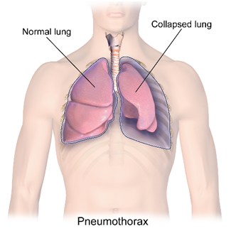
“Pneumothorax” is the medical term for air trapped between lung and ribcage. When air escapes from the lungs it fills the space between the lung and chest wall. As the amount of air in this space increases, the pressure against the lung causes the lung to collapse. This prevents the lung from expanding like it normally would when you breathe in.

What are the types and causes of Pneumothorax?
The 2 different types of Pneumothorax are Non Traumatic/ Spontaneous Pneumothorax and Traumatic Pneumothorax
- Spontaneous Pneumothorax/ Non Traumatic Pneumothorax
This is further classified as follows
Primary Spontaneous Pneumothorax (PSP) – occurs in people who have never been diagnosed with lung disease. The reason for this occurrence is not well understood but established risk factors include males more than females, smoking, and a familial predisposition to pneumothorax
Sometimes small air blisters (small ones are called blebs and larger ones are called bullae) can develop on the top of the lung. It’s uncertain why these blebs appear on some people’s lungs. These blebs rupture and cause pneumothorax.
The causes of rupture of these blebs can be due to change in atmospheric pressure during Scuba diving, Flying, Mountain climbing, Sky Diving.
Secondary Spontaneous Pneumothorax (SSP) – occurs in individuals with significant underlying lung disease. Damaged lung tissue is more likely to collapse.
- Cause – Lung damage can be caused by many types of underlying diseases like
- Chronic obstructive pulmonary disease (COPD) such as emphysema or chronic bronchitis is the cause for almost 70% of SSP.
- Infections of the Lung, such as tuberculosis or pneumonia
- Lung cancer and sarcomas involving the lung.
- Catamenial pneumothorax (associated with the menstrual cycle and sometimes related to endometriosis in the chest)
- Traumatic Pneumothorax (TP)
TP usually occurs after any blunt or penetrating injury to the chest or lung wall. This trauma can damage these structures, and this allows air to leak into the pleural space causing the lung to collapse.
Causes –
- Assault injuries like knife, gunshot wounds
- Accidental injuries like blunt trauma from a car crash or deployment of a vehicle’s air bag
- Fractured ribs, which may puncture the lung
- Iatrogenic injuries like cardiopulmonary resuscitation (CPR), during insertion of chest tubes, lung biopsies taken via a needle through the chest wall
What are the symptoms of a Pneumothorax?
Symptoms may include:
- Chest pain that usually has a sudden onset, sharp and may lead to feeling of tightness in the chest.
- Severe tachycardia, or a fast heart rate
- Rapid breathing or shortness of breath, or dyspnea
- Turning blue, or cyanosis due to decrease in blood oxygen levels.
What is tension Pneumothorax?
A tension pneumothorax is a pneumothorax (primary spontaneous, secondary spontaneous, or traumatic) which results in compression of the chest structures, including vessels that return blood to the heart. Tension pneumothorax can be fatal if not treated immediately.
How is Pneumothorax diagnosed?
History and physical examination – Signs and symptoms following a history or trauma, altitude change or concurrent lung disease can be suggestive of pneumothorax.
Findings on physical lung examination (auscultation) can be varied
- Respiratory distress or respiratory arrest
- Tachypnea
- Asymmetric lung expansion
- Distant or absent breath sounds
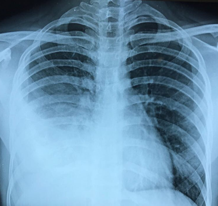
Chest X Ray– The diagnosis of Pneumothorax is confirmed on a Chest X ray. The size of the pneumothorax can also be determined with a reasonable degree of accuracy on the X ray. The size of pneumothorax dictates its treatments upto some extent.

Hydro-pneumothorax (Air + Fluid) seen on the right side with collapsed lung
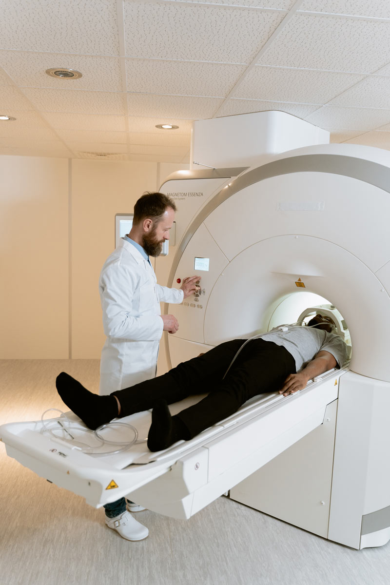
Computed Tomography
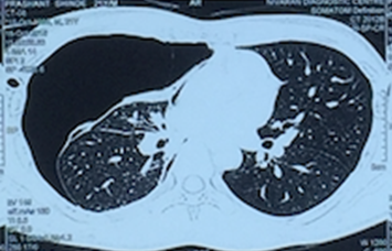
CT scan is used to determine the cause of pneumothorax such as bullae etc. It can also be used to pinpoint the exact size and location of the pneumothorax. In secondary pneumothorax it can also help to identify the underlying lung disease.

Right sided pneumothorax (trapped air) with collapsed lung seen
How is Pneumothorax treated?
The primary aim while treating a pneumothorax is to relieve the pressure on the lung, by removing the air which then allows the lung to re-expand.
Treatment is determined by various factors like
- The severity of symptoms
- The presence of tension pneumothorax
- The presence of underlying lung disease
- The size of the pneumothorax
- Previous history of pneumothorax
- The physical condition of the patient
Based on these the following treatment modalities can be adopted
Observation
Your doctor will keep you under observation if your pneumothorax
- is spontaneous,
- involves only a small area of the chest(<30% of the volume of the hemithorax)
- there is no breathlessness
- no underlying lung disease
He may also take frequent X-rays to check if your lung has expanded again. In such cases sometimes the air gets absorbed from the pleural space. Estimated rates of resorption are between 1.25% and 2.2% of the volume of the cavity per day.
During this period your doctor will instruct you to rest to help healing. Vigorous activity might delay or stop the re-expansion process. Supplemental oxygen may speed the absorption process. Pneumothorax can cause oxygen levels to drop in some people. This condition is called “hypoxemia.” If this is the case, your doctor will order oxygen supplementation along with bed rest.
Secondary pneumothorax istreated conservatively only if the size is very small (1 cm or less air rim) and there are limited or no symptoms.
Needle Aspiration or Chest tube insertion
If a larger area of lung has collapsed, a needle or chest tube will be used to remove the air. These are the two most non invasive methods of treatment of pneumothorax. Local anaesthesia is given to the patient and a hollow needle or tube is inserted between the ribs into the air-filled space. With the needle, a syringe is attached so the doctor can pull out the excess air. Chest tubes are often attached to a mechanical suction device that continuously removes air from the chest cavity and may be left in place for several hours to several days.
Pleurodesis
Pleurodesis is a surgical treatment option and is more invasive than chest tube insertion. Pleura is the membrane surrounding each lung. Pleurodesis is a procedure performed to make your lung membranes stick together so that the pleural space disappears and prevents pneumothorax.
Mechanical pleurodesis is when your surgeon gently rubs the pleura to cause inflammation. Chemical pleurodesis is when your doctor adds chemical irritants to the pleura through a chest tube. During the healing process, the lung adheres to the chest wall, effectively obliterating the pleural space. Recurrence rates are approximately 1%
Bullectomy
In majority of cases of spontaneous pneumothorax, the cause is rupture of a bulla or bleb. These are nothing but air sacs on the lung surface. Larger ones are called as bullae and smaller ones are called blebs. These can rupture due to several reasons such as blunt trauma to the chest, heavy bout of cough, traveling to high altitudes etc. Rupture causes air to leak out of the lungs and accumulate into the chest cavity thereby causing a pneumothorax.
Bullectomy is the surgery for removal (resection) of these bullae. It is usually done by VATS i.e. Video-assisted Thoracoscopic Surgery. Bullectomy is always combined with pleurodesis.
What is the Prognosis?
Your prognosis depends on how quickly your pneumothorax was diagnosed. Early treatment is associated with full recovery. However, in severe cases, late treatment may result in circulatory or respiratory failure. Delay in timely surgery also requires longer recovery. This is often accompanied by worse outcomes, such as incomplete lung expansion and prolonged air leak through the chest tube.
Having one pneumothorax increases the odds for a second. Get medical attention as soon as possible if your symptoms occur again.
Book Appointment

+91-9920024001
