Pleural Effusion
What is Pleural Effusion?
The area between the lining of the lung and the chest wall is called the pleural space. Pleural effusion is the abnormal buildup of fluid in the pleural space. It may also be referred to as effusion or pulmonary effusion.

Normal Lung
What are the types of Pleural Effusion?
There are different types of pleural effusions depending on the nature of content in the pleural space.
- Hydrothorax (Serous Fluid)
- Hemothorax (Blood)
- Chylothorax (Chyle)
- Pyothorax / Empyema (Pus)
- Pneumothorax (air – can be present along with hydro or heamothorax)
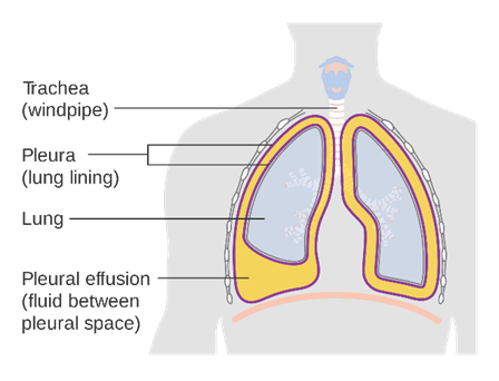
Pleural Effusion (Fluid in the chest cavity)
What are the causes of Pleural Effusion?
A wide range of conditions can cause a pleural effusion. Some of the more common ones are–
- Tuberculosis – this is the most common cause of pleural effusions in developing countries.
- Cancer – usually lung cancer, but any cancer that has progressed to the lung can cause it.
- Infection (empyema secondary to bacterial pneumonia)
- Leakage from other organs: This is usually from heart failure or from liver or kidney disease when fluid builds up in your body and leaks into the pleural space.
- Trauma
- Pulmonary infarction and embolism
What are the symptoms of Pleural Effusion?
Physical symptoms of a pleural effusion commonly show up as liquid fills the chest cavity. These symptoms include:
- Cough
- Chest pain
- Difficulty and pain while breathing
- Fever
- Shortness of breath
- A person who often experience hiccups or a pattern of hiccups that do not go away may also be experiencing pleural effusion.
Some people do not experience symptoms of pleural effusion at all. They usually find out about the liquid in their chest after a physical exam or chest x-ray for an unrelated condition.
How is Pleural Effusion diagnosed?
Medical history and Physical examination – Pleural effusion can be suspected if the patient gives a medical history outlining any or all of the signs and symptoms mentioned above. On physical examination there can be decreased movement of the chest of the affected side, diminished breath sounds, dullness to percussion, fremitus and pleural frictional rub
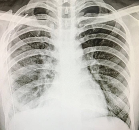
Imaging – The diagnosis is usually confirmed by a Chest X Ray. CT scan is required to further diagnose the cause and extent of pleural effusion.
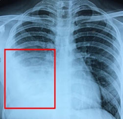
Right sided pleural effusion seen (white area within red box)
How is Pleural Effusion treated?
Draining Fluid
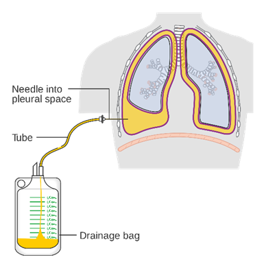
The initial treatment of choice is almost always drainage of the accumulated pleural fluid, either with a needle or a small tube inserted into the chest. You’ll receive a local anesthetic before this procedure, which will make the treatment more comfortable. You may need this treatment more than once if fluid re-collects.
Pleurodesis
Pleurodesis is the process of closing the gap between the pleura of the lung and chest cavity to prevent liquid from building up between them. Your doctor injects an irritating but safe substance (such as talc or doxycycline) through a chest tube into the pleural space. The substance inflames the pleura and chest wall, which then bind tightly to each other as they heal. Pleurodesis can prevent the recurrence of pleural effusions, in many cases.
Surgery
Some pleural effusions may require surgery to break up adhesions, and remove potentially dangerous inflammatory and unhealthy tissue. Pleural effusions that cannot be managed through drainage or pleurodesis may require surgical treatment.
The two types of surgery include:
Video-assisted thoracoscopic surgery (VATS)
A minimally-invasive approach that is completed through 1 to 3 small (approximately ½ -inch) incisions in the chest. Also known as thoracoscopic surgery, this procedure is effective in managing pleural effusions that are difficult to drain or are loculated or recur due to malignancy. Pleurodesis (chemical or mechanical) is done at the end of the surgery to prevent the recurrence of fluid build-up.
Pleural effusions which stay for a long time and/or are infected get converted to empyema. In such cases, surgery is the only treatment of choice for complete recovery and restoration of the lung. In this surgery called as Decortication, your surgeon will peel of the thickened infected layer of pleura from the lung surface so that the lung is able to re-expand completely.
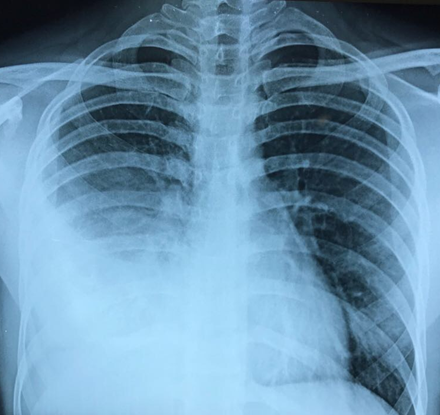
Before
Thoracotomy (Also referred to as traditional, “open” thoracic surgery).
A thoracotomy is performed through a larger incision in the chest. It is required for long standing empyema with very thick pleura, which cannot be removed by the VATS approach. Patients will require chest tubes for few days after surgery to continue draining fluid.
Your surgeon will carefully evaluate you to determine the safest treatment option and will discuss the possible risks and benefits of each treatment option.
After
Book Appointment
+91-9920024001
