
Medically reviewed by Dr Amol Bhanushali, thoracic and chest surgeon | 9 min read
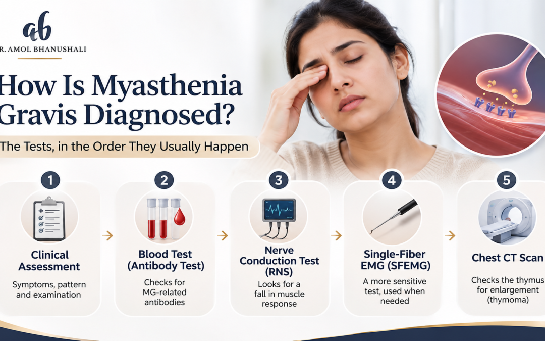
Myasthenia gravis is diagnosed by combining a clinical examination for fatigable muscle weakness with bedside tests like the ice pack test, a blood test for acetylcholine receptor or MuSK antibodies, nerve conduction studies with repetitive stimulation or single-fibre EMG, and a CT or MRI of the chest to check the thymus gland. No single test is enough on its own. The chest scan is where a thoracic surgeon often comes in.
Key insights
- The weakness itself is the first clue: it gets worse with use and better with rest, especially in the eyelids and eyes.
- A negative blood test doesn’t rule it out: around half of people with eye-only myasthenia test negative for the main antibody.
- Single-fibre EMG is the most sensitive test: it picks up abnormal nerve-muscle signalling in more than 95% of patients.
- Everyone diagnosed needs a chest scan: about 1 in 10 people with myasthenia gravis have a thymoma.
Before your tests
Keep a short symptom diary for a week: when your eyelid droops, when you see double, when chewing or climbing stairs gets harder, and whether it’s worse in the evening. Myasthenia often looks normal in a morning clinic, and your diary may be the most useful evidence in the room.
Myasthenia gravis has a habit of hiding. Your eyelid droops by evening and looks fine by morning. You see double while driving home, but not in the doctor’s room. Chewing a meal tires you out, then you’re fine after a rest. It’s easy for everyone, including you, to wonder if it’s real.
It is, and it can be diagnosed with confidence once the right tests are done in the right order. Here’s what each one involves, what it can and can’t tell you, and why a chest specialist in Thane might be asked to look at your scan.
What is myasthenia gravis?
Myasthenia gravis is an autoimmune condition where antibodies disrupt the signal between nerves and muscles, causing weakness that worsens with use and improves with rest. It most often affects the eye muscles, eyelids, face, throat and limbs. It can start at any age.
Who gets it
It’s most common in women aged 20 to 40 and men aged 50 to 80, though it can appear at any age, including childhood.
Where it shows up first
The eyes are affected first in about 40% of people, and eventually in about 85%. Around 15% only ever have eye symptoms, called ocular myasthenia.
Why the thymus matters
The thymus, a gland behind the breastbone, is closely linked to the condition. Many people with myasthenia have an enlarged thymus, and some have a thymoma, a tumour of the thymus.
Test 1: What does the doctor look for in the examination?
The doctor looks for fatigable weakness: a drooping eyelid or double vision that worsens with sustained upward gaze, weakness that worsens with repeated movement, and changes in speech, swallowing or facial expression. Normal reflexes and sensation help distinguish myasthenia from other nerve conditions.
Simple bedside checks
Looking up for a minute or more, holding arms out, counting aloud or repeated squats can bring out the weakness.
What to tell your doctor
When symptoms are worst, whether they change through the day, any choking or breathing difficulty, and any recent infections or new medicines.
Test 2: What is the ice pack test?
The ice pack test involves placing an ice pack over the closed eyelid for about two minutes. If a drooping eyelid lifts noticeably afterwards, the test is positive, suggesting myasthenia gravis. It’s simple, quick and done in the clinic, and it’s about 80% sensitive for eyelid droop.
Why it works
Cooling improves signal transmission at the nerve-muscle junction, temporarily strengthening the eyelid muscle.
Its limits
It’s useful for drooping eyelids but less helpful for double vision. A negative result doesn’t exclude myasthenia.
Test 3: Which blood tests diagnose myasthenia gravis?
The main blood test checks for acetylcholine receptor (AChR) antibodies, which are found in 80% to 90% of people with generalised myasthenia and about 50% with eye-only disease. If that’s negative, doctors test for MuSK antibodies and sometimes LRP4 antibodies.
What the numbers mean
According to the MSD Manual, AChR antibodies are present in 80% to 90% of generalised cases and about 50% of ocular cases, and MuSK antibodies appear in 30% to 60% of patients without AChR antibodies.
Seronegative myasthenia
Some people test negative for all antibodies and still have myasthenia. In them, nerve studies become even more important.
Antibody levels don’t measure severity
A high level doesn’t mean a worse disease. The test is for diagnosis, not tracking how you’re doing.
Test 4: What nerve tests are used?
Two electrical tests are used: repetitive nerve stimulation, which looks for a drop of more than 10% in muscle response with repeated stimulation, and single-fibre EMG, which measures tiny variations in signal timing. Single-fibre EMG is the most sensitive test available.
| Test | What it does | How often it’s abnormal in myasthenia |
| Ice pack test | Cools the eyelid to see if droop improves | About 80% for drooping eyelids |
| AChR antibody | Blood test for the main antibody | 80% to 90% generalised, about 50% ocular |
| MuSK antibody | Blood test if AChR negative | 30% to 60% of AChR-negative patients |
| Repetitive nerve stimulation | Looks for a fall in muscle response | About 60% |
| Single-fibre EMG | Measures signal timing at single fibres | More than 95% |
Why single-fibre EMG isn’t first
It’s highly sensitive but not specific, because other nerve and muscle conditions can also cause abnormal results. It’s best used alongside the clinical picture.
Test 5: Why do you need a chest CT or MRI?
Everyone diagnosed with myasthenia gravis should have a CT or MRI of the chest to look at the thymus gland. About 10% of patients have a thymoma, and many more have an enlarged thymus. Finding either changes treatment and brings a thoracic surgeon into the picture.
Thymoma vs chest wall tumour
A thymoma sits in the middle of the chest, behind the breastbone, so it usually isn’t something you can feel. That’s quite different from chest wall tumor symptoms, which often include a lump you can see or feel. Thymomas are usually found on a scan, sometimes causing chest pain, cough or breathlessness if large.
What happens if a thymoma is found
A thymoma is usually removed surgically, whether or not you have myasthenia. About half of thymomas are malignant, so complete removal matters.
Test 6: What other tests might you need?
Doctors often check thyroid function, because thyroid disease is more common in people with myasthenia, and breathing tests like forced vital capacity, to check whether breathing muscles are affected. Other tests depend on symptoms and treatment plans.
Breathing tests matter
Myasthenic crisis, where breathing muscles weaken dangerously, affects around 15% to 20% of patients at some point. Baseline breathing measurements help spot trouble early.
Where does surgery fit in after diagnosis?
Surgery to remove the thymus, called thymectomy, is recommended for anyone with a thymoma and is also offered to many people with generalised AChR-antibody-positive myasthenia without a thymoma. It can reduce symptoms and the need for long-term steroids.
What the trial showed
The MGTX trial randomised 126 patients with generalised, non-thymoma myasthenia to thymectomy plus prednisone or prednisone alone. Over three years, the surgery group had better symptom scores, needed less prednisone and were hospitalised for exacerbations less often, 9% against 37%.
Keyhole or open?
Thymectomy can be done through the breastbone or by minimally invasive keyhole techniques such as VATS, depending on the case and whether a thymoma is present.
Teamwork
Diagnosis and medical treatment are led by a neurologist. The thoracic surgeon reviews the chest scan, advises on thymectomy and performs it.
Wrapping up
So how is myasthenia gravis diagnosed? Through a careful examination, bedside tests like the ice pack test, antibody blood tests, nerve studies and a chest CT or MRI to check the thymus. No single test is decisive, and a negative antibody test doesn’t rule it out. Once it’s confirmed, the chest scan decides whether surgery to remove the thymus belongs in your plan.
Dr Amol Bhanushali, a chest specialist in Thane trained at Tata Memorial Hospital, works with neurologists to treat thymoma and myasthenia gravis with thymectomy, including minimally invasive approaches where suitable. If you’ve been diagnosed and your scan shows thymus changes, or you’ve been told you might need thymectomy, book a consultation to understand your options.
FAQs
What is the first test for myasthenia gravis?
A clinical examination looking for fatigable weakness, often with a bedside ice pack test for drooping eyelids. A blood test for acetylcholine receptor antibodies is usually ordered next to support the diagnosis.
Can you have myasthenia gravis with a negative blood test?
Yes. About half of people with eye-only myasthenia and some with generalised disease test negative for AChR antibodies. MuSK and LRP4 antibody tests and single-fibre EMG help in these cases.
What is the most accurate test for myasthenia gravis?
Single-fibre EMG is the most sensitive, detecting abnormalities in more than 95% of patients. It isn’t specific to myasthenia, so it’s interpreted alongside the examination and antibody results. Your neurologist reads all three together.
Why do you need a CT scan for myasthenia gravis?
To check the thymus gland. About 10% of patients have a thymoma, and many have an enlarged thymus. Finding either affects treatment, especially whether thymectomy is recommended. An MRI can be used instead.
Does myasthenia gravis need surgery?
Not always. Thymectomy is recommended if there’s a thymoma and is offered to many people with generalised AChR-positive myasthenia. A trial showed it improved symptoms and reduced steroid needs over three years.
Who diagnoses myasthenia gravis?
A neurologist usually leads the diagnosis and medical treatment. A thoracic surgeon becomes involved when the chest scan shows thymus changes or thymectomy is being considered. The two specialists usually work as one team.